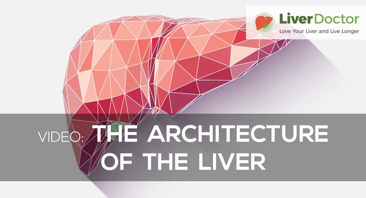
The Architecture Of The Liver
The liver is the largest and hardest working organ in the body. Liver tissue is composed of a compact mass of multi-sided units known as the hepatic lobules. Each lobule has a central vein, which joins the hepatic vein which joins a huge vein known as the inferior vena cava and takes blood back to the right side of the heart; this blood returning to the heart has been processed and cleansed in the liver lobule. Each lobule has layers and rows of liver cells (hepatocytes) which do all the detoxification of the blood as it flows through the lobule.
The liver receives blood from two sources which is unique to the body's organs. Eighty percent of the liver's blood supply arrives in the portal vein, which carries blood from the intestines and is high in digested food materials and toxins from the gut. The remaining twenty percent of the liver's blood supply arrives in the hepatic artery and contains oxygenated blood from the heart. Obviously the liver cells need a lot of oxygen to perform their metabolic functions.
Sinusoids are the spaces between plates of liver cells and tiny vessels off the end of the hepatic artery and portal vein. The sinusoids conduct the blood flow to the central vein. Inside the spaces (sinusoids) an exchange of materials/chemicals takes place between the liver cells and the blood; this enables nutrients/toxins to be removed from the blood.
Only after the blood is cleansed does it pass into the central veins which return to the hepatic vein which returns the blood to the general body circulation via the huge vein called the vena cava.
The liver cells manufacture bile and pump it into tiny bile canaliculi (seen in green) which carry the bile to branches of the bile duct which convey bile to the gall bladder.
When an adult is at rest, about two and a half pints of blood flow through the liver each minute.
Problems with liver architecture can be visualised on an ultrasound scan of the liver as well as a CT or MRI scan of the liver.
Know someone who might benefit from this article? Share it!


2 Comments
Search
Shop Our Products by
Health Interest
-

Hormone & Thyroid Health
-

Liver & Gallbladder Health
-

Digestive Health
-

Bone & Joint Health
-

Women's Health
-

Immune Health
-

Healthy Aging
-

General Health
Most Recent Articles
Need Help?
1-888-75-LIVER
Monday to Friday, 9:00 am to 5:00 pm MST
100%
Satisfaction Guaranteed
If it’s faulty or wrongly described, we’ll replace it.
Hepatocytes are the main cells of the liver.
Kind regards,
Jessah Shaw
Nutritionist